September 9, 2024
How To Remove Cherry Angiomas
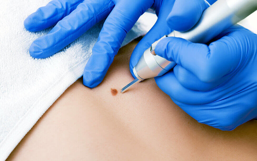
Cherry Hemangioma Problem, Therapies And Images For Adults Cut excision permits fragile elimination of the lesion by blade and histologic verification of the diagnosis. Hemostasis complying with elimination may be acquired by chemical means (aluminum chloride) or by executing electrocautery. After numbing the skin, the angioma is slashed off and sent for pathological exam. This approach is recommended if there's a suspicion of change or malignancy in the angioma. They are not hazardous and normally do not cause any kind of signs. Nonetheless, they might bleed or itch sometimes, as stated earlier.
If You Observe Changes In Previously Steady Skin Lesions
Older Than 30? You Might Start Noticing These Moles on Your Skin - Well+Good
Older Than 30? You Might Start Noticing These Moles on Your Skin.
Posted: Mon, 22 Apr 2024 07:00:00 GMT [source]
Cherry angiomas are usually gotten rid of for aesthetic factors, as cherry angiomas are not an indication of any kind of hidden wellness issues or medical conditions. Many people who have cherry angiomas may pick to have them gotten rid of if they are dissatisfied with their cosmetic look. There are times, nonetheless, when the removal of a cherry angioma is required as a result of inflammation or constant blood loss. Effective treatment options for cherry angiomas include the V-Beam laser, cryosurgery, cut excision, IPL, and electrodesiccation.
What Medical Conditions Are Cherry Angiomas Linked To?
The biopsy treatment may be made use of as a healing procedure to eliminate shocked or bleeding lesions. Cherry moles, or angiomas (also commonly referred to as "cherry angiomas"), are red moles-- normally benign-- which contain tiny capillary that give them their shade. Cherry angiomas are so called as a result of the collection of tiny blood vessels which give them a reddish appearance.
Surgical Elimination
Cherry angiomas and cherry hemangiomas are common, benign skin growths that are extremely similar in appearance however are made from various cells. While cherry angiomas are constructed from lymphatic or blood vessels, cherry hemangiomas are made from only blood vessels. Cherry hemangiomas might appear in infancy or childhood years, whereas cherry angiomas normally impact adults.
- Some people additionally describe cherry angiomas as senile angiomas, capillary angioma, cherry hemangioma, Campbell de Morgan spots or simply cherry red skin papules/moles.
- If you do choose to have them dealt with, electrodessication, fluid nitrogen, or laser treatment might be used by your skin specialist.
- Prior to the procedure, the location is numbed with a shot of local anesthetic.
- They can occur nearly anywhere on the body, and the majority of commonly begin showing up around age 40.
- Well, cherry angiomas are the most common type of angiomas, or benign lumps, adults develop on their skin.
- Any individual who wants to go through a cherry angioma elimination for aesthetic reasons need to consult their medical professional to talk about the alternatives.
The specific reason for red moles is unknown, but there may be a hereditary aspect that ensures individuals more likely to get them. They've additionally been connected to maternity, exposure to chemicals, specific medical problems, and environment. To learn more concerning cherry angioma/hemangioma treatment at HMGS Dermatology, get in touch with our workplace today to schedule an appointment. Cherry angiomas commonly provide in the 3rd or fourth years of life, and very early sores might look like tiny red macules. Store the mix at room temperature level and apply it to the skin several times daily. This can additionally be made use of on the scalp and face in most individuals without side effects, along with for various other skin conditions. Genes contribute in just how likely you are to have cherry angiomas. If your parents and grandparents have them, there's a great chance you will, as well. Direct exposure to particular chemicals and gases in the environment can also trigger cherry angiomas to show up in clusters. Nevertheless, cherry angiomas can range in shade and may additionally look blue or purple. If pressure is put on them, they may for a moment transform white. A hemangioma commonly appears on the face, scalp, chest or back, though it can be anywhere on the skin. Therapy usually isn't required for a child's hemangioma, as the mark fades gradually. You might wish to think about treatment for the kid if a hemangioma brings about problems with vision, breathing or other physical features. You additionally may think about therapy if the hemangioma is in a cosmetically sensitive area. This might make its appearance even worse and might cause scarring. If you wish to remove a cherry angioma, it is best to look for expert help. Age might also
https://nyc3.digitaloceanspaces.com/5ghb9bmaj7etny/Wellness-service/mole-removal/over-the-counter-medical-treatments-for-typical.html be a factor in cherry angiomas as they generally begin around age 30 and appear to grow in size with age. Cherry angiomas are commonly small, bright red, and circular or oval-shaped, varying in between the dimension of an identify to as much as a quarter-inch in diameter. They typically are found on your torso, arms, legs, or shoulders.
Do cherry angiomas hemorrhage a great deal?